UPPER CERVICAL CHIROPRACTIC CARE PART 3: The Images
In Part 3 of our Upper Cervical Chiropractic Care series we’re diving into another key component of what makes Upper Cervical Chiropractic unique; the specialist images of your spine that give us the precise insights into the adjustment/s that your body needs.
A summary of what we’ll cover:
> Special Images to Understand Your Spine and Body Better
> What is the Bio-mechanical Series of Images?
> What Important Information Do These Bio-mechanical Images Give Us?
> What Does The Lateral View of Your Neck Look Like? What Does It Show Us?
> What to expect when you get your images taken at our partner imaging centre
> What About Children? Why We Don’t Usually Need Images for Children
Recap:
> PART 1: The difference between regular Chiropractic and Upper Cervical
> PART 2: The Scanner - Computerized Infrared Thermography
Up next:
> PART 5: What to expect in your Upper Cervical Chiropractic care program
Special Images to Understand Your Spine and Body Better
As part of your new client process we will assess if its necessary to take specialist images of your upper cervical spine. For all clients that we believe have a problem in their upper cervical region, what is officially called a vertebral subluxation, we’ll request they get these special images taken.
There are 4 images that are taken as standard for upper cervical chiropractic. Three of these images are referred to as a bio-mechanical series. The fourth image is simply a view of your neck from the side, called a lateral view. This image is a common view usually taken by regular doctors and chiropractors, but the bio-mechanical series is a special set of views only chiropractors trained in Knee Chest Upper Cervical use.
SIDE NOTE: If you’re reading this blog, and your UC Chiro practices a different UC technique to Knee Chest, its highly likely they’ll take a different series of views (they may also do it in-house as chiropractors are actually trained how to take and analyse radiographs. This is just not permitted in Spain, hence why we’ve partnered with a great imaging centre). Whatever the views taken by your UC Chiro, the objective is always the same; we want to see what’s happening with your Atlas and Axis in relation to your skull and your lower neck bones.
Another important side note is that in other countries like the USA or UK it may be possible that your UC Chiro has a special technology called 3D CBCT which is a 3D image taken by a moving machine to see your upper cervical spinal bones.
What is the Bio-mechanical Series of Images?
Each of the three images are taken with your mouth open, so that we can see your Atlas and Axis.
The first image is of you with your head straight on. The other two images are taken with you side-bending your head towards your ear, one to the left and one to the right.
Inside your mouth is where we see your Atlas and Axis, their position, their relationship to your skull and their range of movement when you side bend. We’ll explain why this is important in just a moment.
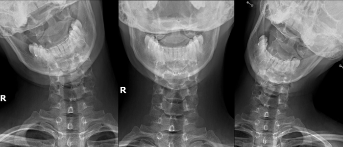
What Important Information Do These Bio-mechanical Images Give Us?
Confirm if you have a problem, called a vertebral subluxation in your Atlas or Axis vertebrae. If so, Upper Cervical care will benefit you.
Help us to determine which vertebra is a priority to adjust, and more specifically in which direction. The adjustment you receive is a precision adjustment, specific to your body, measured from your radiograph images and not just based on what we ‘feel’ on the outside.
What Does The Lateral View of Your Neck Look Like? What Does It Show Us?
This is a standard view that’s commonly taken by traditional medical doctors and chiropractors. However, what we are analysing it for is different to a medical doctor.
A medical doctor prioritises making sure that you have no pathological issues, especially following a trauma (i.e. making sure there are no fractures) and also checking the adjacent soft tissue structure, and if you have osteoarthritis (thinning of the bone) or spondylosis (what doctors consider wear and tear of your neck bones).
Many times clients have had this image taken and assessed by a traumatologist or other doctor and told everything is ‘normal’ or they have wear and tear which is ‘expected for their age.’
This can feel frustrating when you are suffering and the doctors can’t seem to find a problem. But maybe they just weren’t looking in the right place.
When we assess this lateral view from a chiropractic point of view we are also interested in the alignment of your cervical spine. And we view the changes from a different perspective.
When assessing the spacing between each vertebrae, and the shape and health of each vertebrae we are not viewing changes as ‘wear and tear’. We understand that your body will make appropriate adaptations in your best interest.
Usually, as the spinal alignment changes, your head moves forwards meaning that it weighs relatively more than it should. This extra strain on your neck bones leads them to make changes (adaptations).
They grow more bone to become stronger to hold the weight. They also move closer together and widen, again to be more stable.
This is not your body failing. This is an awesome, smart response of your body!
If we see lots of adaptation or just a little adaptation it allows us to have a better understanding on how hard your body has been working and therefore how easily it may return to normal when it no longer needs to adapt so much.
We also use it to get to know the size, angle and position of your atlas and axis bones from the side. This helps us in our ability to deliver your specific adjustment in the precise place that it needs to be made.
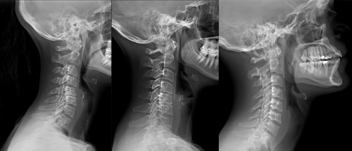
Examples of different people’s necks in lateral view and what they mean.
Left: This image is showing a lot of adaptation that has probably taken a long time to occur. Alternatively this person may have suffered a big physical trauma that resulted in a big change, quickly. Somebody with a neck like this may take longer to change and return to normal function. They might also notice more ups and downs in their journey as their body has been adapting harder, and potentially for longer.
Centre: This person has less adaptation occurring that the left image, however there is still a fair amount of adaptation.
Right: This is an example of a body that has had to make less adaptations. Its closer to a normal neck. Generally (of course nothing is ever set in stone as each human is a different world, but to give you an idea) somebody with this type of neck will typically respond faster and hold their adjustments easier.
What to Expect When You Get Your Radiographs Taken at our Partner Imaging Centre
Firstly, it’s important to know that in the UK (as well as the USA, Australia and Canada) Chiropractors are trained and authorised to take their own radiographs. Many Chiropractors have the machines in their offices to be able to do this, or they may refer to another local Chiropractor. When I worked in the UK (from 2010-2016) I took the radiograph images myself within the office.
However in Spain, Chiropractors are not allowed to take their own radiographs.
I have therefore partnered with Creu Blanca on Carrer de Corsega and trained their head radiologist on how to take the special bio-mechanical series. The team are there Monday to Friday, 8am-8pm and can take your images in the moment without the need for you to book an appointment.
For you to be able to get these images taken I will give you two papers:
A request form which tells Creu Blanca we want these special pictures taken of your cervical spine
A permission form that you sign, which allows the centre to email me the access to your images. They usually do this around 4 hours after they take the images.
When you arrive at the centre they will simply send you downstairs to the ‘Radiografias’ department. The receptionist will check you in. As soon as they are available one of the technicians will take you through to an imaging suite. You’ll need to take off your earrings and necklaces, and potentially put on a gown (depending on what clothes you are wearing). They’ll run through the 4 images and once you’ve finished and changed, you’ll head back out to reception to pay. The total cost for the 4 images is 70€ at the time of writing. If you have private medical insurance it may cover the cost but its important that you speak with them before you go incase you need a special authorisation from your insurance company (each company and policy is different).
Once I have received access to view your images online I will be able to analyse them in preparation for your first adjustment.
What About Children? Why We Don’t Usually Need Images for Children
For most children, we do not need to take radiograph images. Firstly we want to avoid exposing them to radiation unless absolutely necessary. Secondly, due to the fact that they have been adapting for way less time than adults, their patterns are usually much more straightforward. This means that it’s normally possible to establish where their specific adjustment needs to be made without the need of a radiograph image.
If your child has had a big trauma, or is under care and isn’t improving then we may discuss the need for a radiograph at that time.
RELATED POSTS


